Clinical Applications for AI Epic™ Co-Ablation System- Liver Cancer

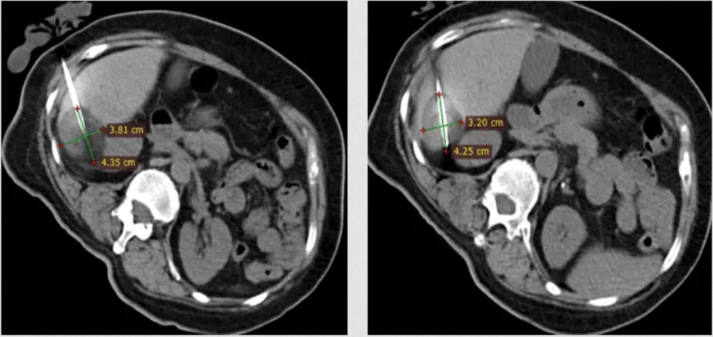
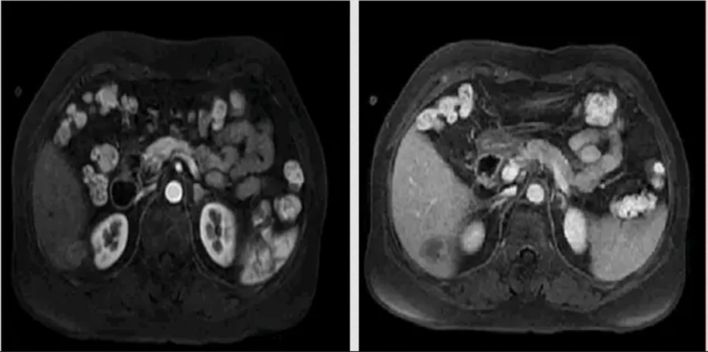
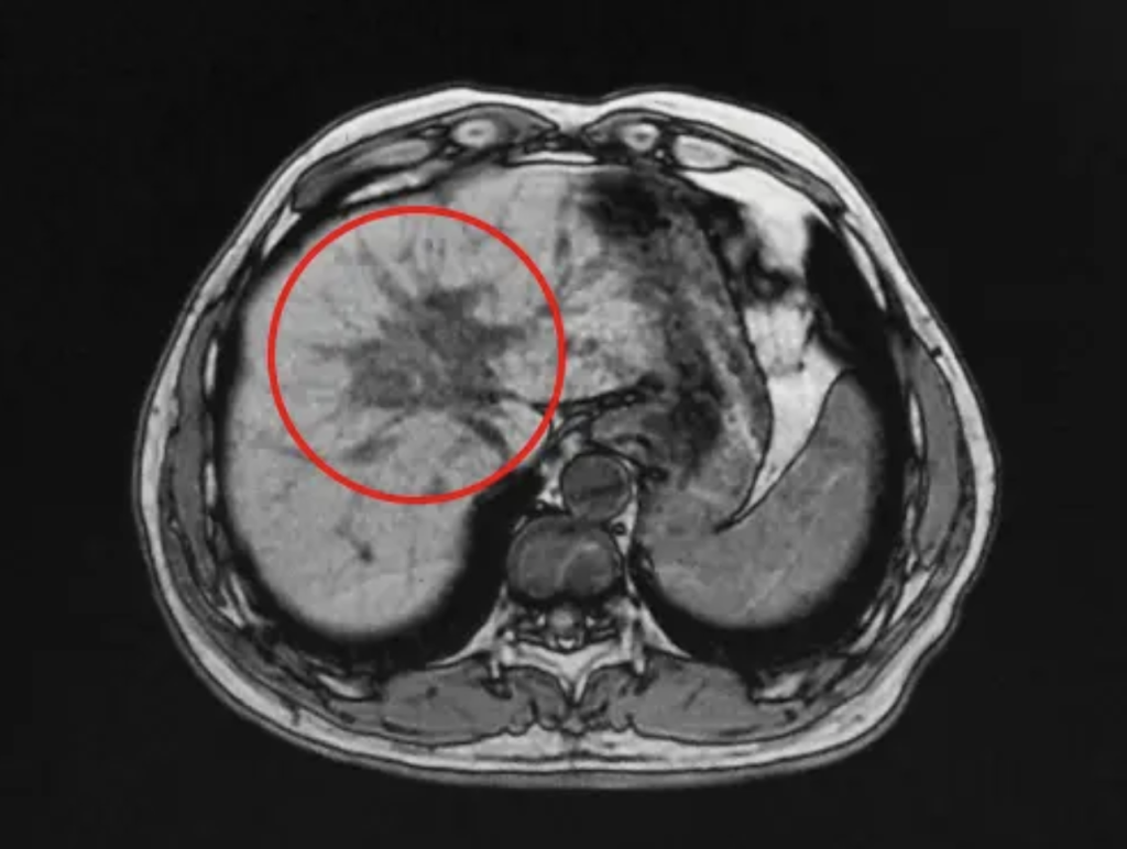
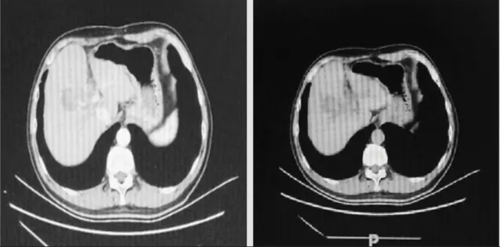
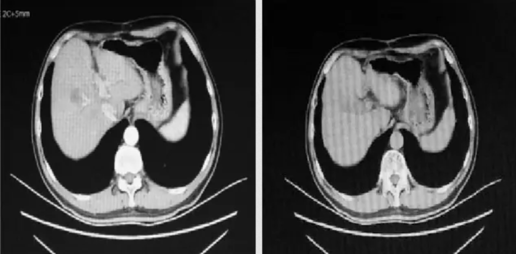
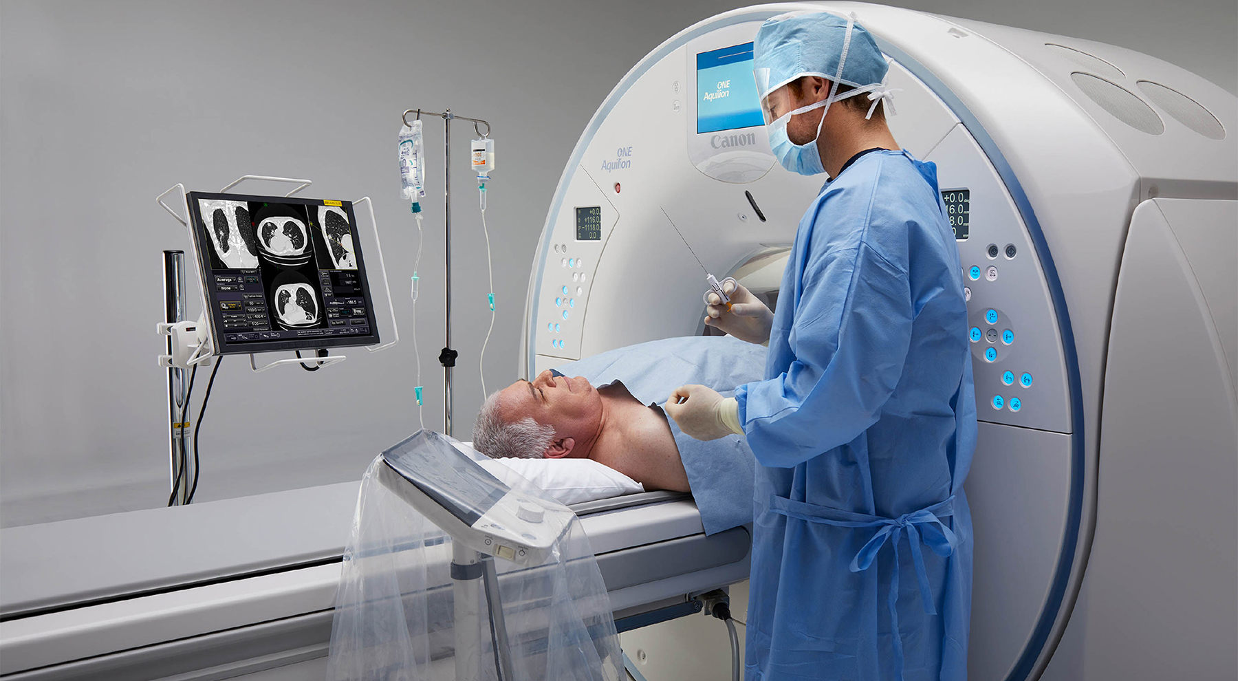
What is Cryotherapy for Liver Cancer? Liver cancer cryoablation is a minimally invasive therapeutic technique that offers a promising treatment option for select liver cancer cases. It utilizes extreme cold temperatures to destroy cancerous cells within the liver while sparing as much healthy tissue as possible. This approach is particularly suitable for patients who are not candidates for surgical resection or conventional therapies. During the procedure, a thin, needle-like probe is precisely inserted into the liver tumor. The probe delivers ultra-low temperatures, typically below -40°C, creating an ice ball that engulfs and destroys the cancer cells. Repeated freezing and thawing cycles induce cellular damage, ultimately resulting in targeted tumor necrosis.

Brand:

HYGEA

Date:

Advantages of Cryosurgery for Liver Cancer

One key advantage of cryoablation is its ability to precisely target localized tumors, especially those not easily accessible through surgery or in patients with multiple small lesions. Additionally, cryoablation is generally well-tolerated and can often be performed on an outpatient basis.

The success of liver cryoablation depends on factors such as tumor size and location. Smaller tumors near the liver surface typically respond better to cryoablation, while larger or centrally located tumors near major vessels may require combination therapies or alternative approaches.

Precise Ablation Protecting Normal Tissue

Cryoablation allows real-time CT or ultrasound imaging to precisely control ice ball formation, targeting the tumor while preserving healthy liver tissue and vital vessels, minimizing collateral damage.

Stimulates Immune Response and Enables Innovative Combination Therapies

Cryoablation releases abundant tumor antigens, activating the immune system and enhancing immune surveillance. It can be combined with chemotherapy, immunotherapy, and other treatments to boost efficacy and improve long-term tumor control.

Minimally Invasive, Safe, Fast Recovery, and Repeatable Treatment

Minimally invasive and safe, cryoablation enables fast recovery with fewer complications. Ideal for patients unsuitable for major surgery, it can be repeated flexibly upon tumor recurrence, expanding treatment options.

Clinical research has demonstrated that liver cryoablation can provide effective local tumor control and potentially improve survival outcomes, particularly when integrated into a multimodal treatment plan. However, as with any medical procedure, there are inherent risks, including bleeding, infection, or potential injury to adjacent structures.

As technological innovations and oncology research continue to advance, liver cancer cryoablation—supported by state-of-the-art cryosurgical systems—is poised to play an increasingly important role in comprehensive liver cancer management. Its minimally invasive nature and ability to address challenging cases position cryoablation as a valuable tool in liver cancer management.

References

1. Guan, H., et al. (2023). Short-term efficacy and safety of CT-guided percutaneous compound cryoablation for liver malignancies. Chinese Journal of Interventional Imaging and Therapy, 20(4), 198–203. 2. Wang, C. et al., Cryoablation for liver cancer, Cancer Letters, 2019. 3. Niu, L. Z., et al. (2014). Percutaneous cryoablation for liver cancer. Journal of Clinical and Translational Hepatology, 2(3), 182–188. 4. Chen, Y., et al. (2022). Cryoablation combined with PD-1 inhibitors for advanced solid tumors: A retrospective study. Frontiers in Immunology, 13, 966008.

Cases of Cryoablation for Liver Cancer

Case #1

Case Characteristics

The lesion was under the liver capsule in segment VI and adjacent to the right kidney. Preoperative CT imaging showed the targeted lesion in the right hepatic lobe, measuring 30.4 × 31.5 mm.

Ablation-two cycles

(freezing for 20min + heating re-warming for 5min)

Re-examination 1 month after surgery

Re-examination 6 months after surgery, MRI showed complete remission (CR)

Case #2

Case Characteristics

The lesion was adjacent to portal vein. CT scan showed that the lesion was at the junction of IV and V segments, sized as 41×32mm.

Ablation-two cycles

(freezing for 20min + heating re-warming for 6min)

Re-examination 1 month after surgery

Re-examination 3 months after surgery, CT showed complete remission (CR)